Allan D, Tieu A, Lalu M, Burger D. Mesenchymal stromal cell-derived extracellular vesicles for regenerative therapy and immune modulation: Progress and challenges toward clinical application. Stem Cells Transl Med 2020; 9(1):39-46.
Rohde E, Pachler K, Gimona M. Manufacturing and characterization of extracellular vesicles from umbilical cord-derived mesenchymal stromal cells for clinical testing. Cytotherapy 2019; 21(6):581-592.
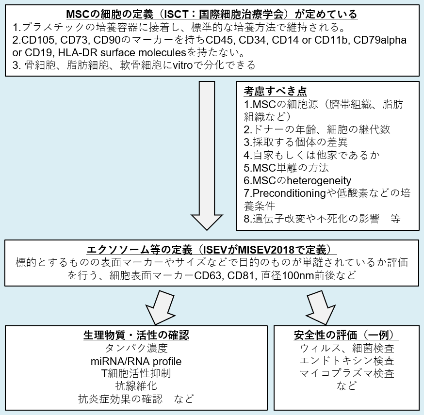
Witwer KW, Van Balkom BWM, Bruno S, Choo A, Dominici M, Gimona M, et al. Defining mesenchymal stromal cell (MSC)-derived small extracellular vesicles for therapeutic applications. J Extracell Vesicles 2019; 8(1):1609206
Elahi FM, Farwell DG, Nolta JA, Anderson JD. Preclinical translation of exosomes derived from mesenchymal stem/stromal cells. Stem Cells 2020; 38(1):15-21.
Tsuchiya A, Takeuchi S, Watanabe T, Yoshida T, Nojiri S, Ogawa M, et al. Mesenchymal stem cell therapies for liver cirrhosis: MSCs as “conducting cells” for improvement of liver fibrosis and regeneration. Inflamm Regen 2019; 39:18.
Lener T,Gimona M, Aigner L, Borger V, Buzas E, Camussi G, et al. Applying extracellular vesicles based therapeutics in clinical trials -an ISEV position paper. J Extracell Vesicles 2015; 4:30087.
Thery C, Witwer KW, Aikawa E, Alcaraz MJ, Anderson JD, Andriantsitohaina R, et al. Minimal information for studies of extracellular vesicles 2018 (MISEV2018): a position statement of the International Society for Extracellular Vesicles and update of the MISEV2014 guidelines. J Extracell Vesicles 2018; 7(1):1535750.
Dominici M, Le Blanc K, Mueller I, Slaper-Cortenbach I, Marini F, Krause D, et al. Minimal criteria for defining multipotent mesenchymal stromal cells. The International Society for Cellular Therapy position statement. Cytotherapy 2006; 8(4):315-317
DMに対するExsm臨床治験
NCT03106246
Circulating Extracellular Vesicles Released by Human Islets of Langerhans
McGill University Health Center
CHU de Quebec-Universite Laval
NCT03660683
Effect of Saxagliptin and Dapagliflozin on Endothelial Progenitor Cell in Patients With Type 2 Diabetes Mellitus
Sabyasachi Sen|George
Washington University
NCT03250078
A Pancreatic Cancer Screening Study in High Risk Individuals Including Those With New-Onset Diabetes Mellitus
Western Connecticut Health
Network
NCT02649465
SGLT2 Inhibitor Versus Sulfonylurea on Type 2 Diabetes With NAFLD
Kanazawa University
Kowa Company, Ltd.
NCT02138331
Effect of Microvesicles and Exosomes Therapy on ホイ-cell Mass in Type I Diabetes Mellitus (T1DM)
General Committee of Teaching Hospitals and Institutes, Egypt
NCT03027726
Prevention of Diabetes in Overweight/Obese Preadolescent Children
Basque Country University
Ministerio de Competitividad, Spain
NCT03392441
Insulin Deprivation on Brain Structure and Function in Humans With Type 1 Diabetes
Mayo Clinic
Obesityに対するExsom臨床治験
NCT03459703
Effect of Time-Restricted Feeding on Fat Loss and Cardiometabolic Risk Factors in Overweight Adults
University of Alabama at Birmingham
NCT03762629
Exercise and Diet Restriction on Cardiovascular Function in Obese Children and Adolescents